what is congenital glaucoma? Babies and young children (under the age of three) have been found to develop congenital glaucoma, also known as childhood glaucoma, infantile glaucoma, or pediatric glaucoma. Despite being a rare condition, it may cause a permanent loss of vision.
What Is Congenital Glaucoma?
The more than a million nerve fibers that make up the optic nerve, a nerve cord that connects the retina to the brain, form the optic nerve. The optic nerve must be healthy in order to guarantee clear vision. The term “glaucoma” refers to a group of ocular conditions that gradually weaken the optic nerve. If the damage is not repaired, it may lead to a smaller visual field or even total blindness.
Even though it is common in adults, some types of glaucoma, like congenital glaucoma, only affect children. One out of every 30,000 live births is affected by this extremely uncommon condition, but it can result in severe and irreversible vision loss in affected children.
What Affects Your Eye As A Result?
A healthy eye has fluid that circulates under normal pressure and draws nutrients in. A network of cells and tissue is traversed by it as it drains. Your eye constantly produces more to make up for what is lost. This process deviates when PCG is used. Most of the time, the fluid doesn’t drain as it should, and the buildup causes your eye pressure to increase.
A signal is sent to your brain by the optic nerve at the back of your eye. The PCG-induced increase in pressure harms the nerve fibers that make up this nerve.
This harm accrues over time with the majority of glaucoma types. When you start experiencing symptoms, oftentimes the damage has already been done. Once your vision is lost, it is impossible to regain it.
What Triggers Congenital Glaucoma?
The anterior chamber is a location inside the eye. Aqueous humour, a clear liquid that fills this area and maintains the eye’s optical properties, bathes the eye’s structural components. The anterior chamber experiences constant entry and exit. It exits through the iris-cornea angle. This angle’s job is to keep the intraocular pressure stable and prevent damage to the optic nerve by allowing the aqueous humour to flow out of the eye. The angle must therefore stay open in order for the aqueous humour to drain from the eye.
Due to a birth defect in the eye’s angle of development brought on by poor eye development, congenital glaucoma causes an increase in intraocular pressure. The result is an increase in intraocular pressure and damage to the optic nerve because the aqueous humour cannot drain normally.
Signs Of Congenital Glaucoma
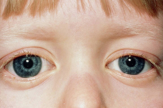
Clinically, elevated IOP >21 mmHg, corneal edema, enlargement of the eye with buphthalmos, and Haab striae are the main indicators of PCG. IOP levels at presentations typically range from 30 to 40 mmHg, though they occasionally fall outside of this range. = If the optic nerve is healthy and the patient’s eye growth is within normal ranges, IOP in the low-20s mmHg is acceptable. However, if there are other more severe signs of PCG, it might not be.
The cornea becomes cloudy as a result of diffuse and/or focal edema when IOP is in the 30–40 mmHg range. In an eye with increased IOP, the endothelial cell layer cannot remove fluid from the cornea as it can in adult eyes. However, in young children, there is the additional insult of corneal stretching from high IOP, which results in Descemet breaks and striae, which are areas of bare stroma bordered by two separated edges of Descemet membrane that become ridges as a result of hyaline deposition. When the IOP is high, these are known as Haab striae and are connected to acute overlying focal corneal edema. Around 25% of PCG eyes that present at birth have them, and more than 60% of PCG eyes are discovered by the time a child is 6 months old. They can be single or multiple, horizontal or oblique in orientation. Corneal edema might go away once IOP returns to normal, but Haab striae might still be present and connected to corneal scarring. Even after the IOP is under control, the poorly controlled cases of PCG may develop dense stromal opacification.
The typical diameter of the cornea in a newborn is between 9.5 and 10.5 mm, and by age 1, it is between 10.0 and 11.5 mm. Any diameter greater than 12.0 mm before the age of one suggests an anomaly, particularly if there is an imbalance between the two eyes. Glaucoma suspicion should be high if the diameter is greater than 13 mm at any age. In the presence of elevated IOP, all eye tissues, including the cornea, the scleral wall, and the lens, are stretched, resulting in buphthalmos. While the sclera can continue to stretch up until age 10, the growth of the cornea stops around the age of three.
Other symptoms of eye distension include an abnormally deep anterior chamber, myopia (mainly because of eye lengthening and enlargement), astigmatism (caused by corneal stretching and Haab striae), anisometropia (almost always present in unilateral PCG), and optic nerve cupping.
Young children’s optic nerve cupping may only be caused by the posterior bowing of the lamina cribrosa and the stretching of the optic canal, without any loss of the neuroretinal rim. A noticeable cupping reversal may occur when the IOP is normalized. The damage to the retinal nerve fiber layer, if it exists, is permanent, whereas cupping may resolve. Cupping develops as a result of neuroretinal rim tissue loss, particularly at the vertical disk poles, in older children and people with advanced glaucoma.
Any asymmetry between the eyes in those glaucoma symptoms should be suspected. The other symptoms mentioned above may also be accompanied by amblyopia, either deprivation or both.

How Is It Diagnosed?
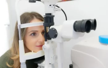
When a baby or child is under three years old, a full eye exam is typically performed in the operating room while the patient is sedated to detect congenital glaucoma.
The test consists of:
- the front of the eye is examined: This aims to evaluate the state of the cornea and the angle before deciding the best surgical method for each case.
- The fundus is examined: The ophthalmologist uses eye drops to dilate the pupils before inspecting the retina and optic nerve with a specialized microscope to check for any damage. The optic nerve’s nerve fibers are gradually lost as a result of glaucoma, leaving an opening (excavation) that widens as the disease progresses.
Congenital Glaucoma Symptoms
Among the symptoms and warning signs of juvenile glaucoma are:
- Triad
- Overflow of tears onto the face (Epiphora),
- Involuntary twitching of the eye (Blepharospasm),
- Sensitivity towards the light (Photosensitivity)
- Enlargement of the eyes (Buphthalmos)
- Hazy cornea
- Closing of the eyelid
- Redness of the eye
Congenital Glaucoma Treatment
Almost always, surgery is the best option. Doctors prefer to administer anesthesia as soon as the diagnosis is confirmed because doing so with young children is risky. A simultaneous operation will be performed on both eyes if both are impacted.
The doctor may recommend eye drops, oral medication, or a combination of the two to help control fluid pressure if immediate surgery is not an option.
Microsurgery is a procedure that many medical professionals perform. To make a canal for the extra fluid to drain, they use small tools. To drain fluid from the eye, the doctor may occasionally implant a valve or tiny tube.
The doctor may use laser surgery to remove the fluid-producing tissue if the conventional methods don’t work. After surgery, they might recommend medication to help control eye pressure.